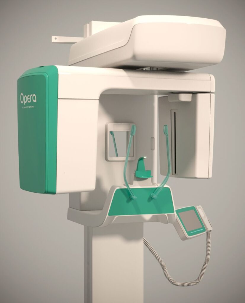
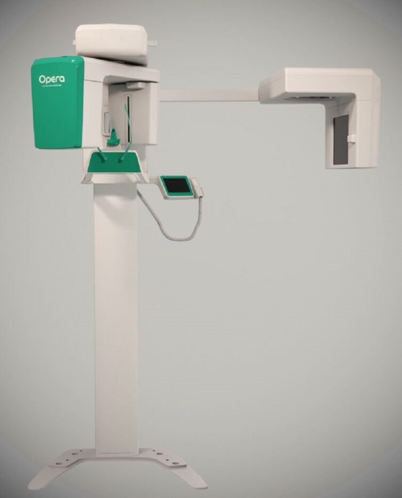
The OPERA family begins with OPERA 2D: thanks to its clean and ergonomic design, OPERA 2D perfectly fits into your workspace and offers high-level technological development for the acquisition of clear 2D images:
• Exposure time of 14.3s for children and 15s for adults
• DC high frequency generator
• 151 x 6.9mm Active Area
• 15 x 30cm HD images
• 0.5mm focal spot
• Sensor TDI CCD
– the standard configuration, not Upgradable, which remains original in its PAN functions;
– the Upgradable version, which is adapted to allow the evolution to 3D and/or CEPH models.
TECHNICAL SPECIFICATIONS FOR OPERA 2D:
Sensor Type PAN CCD, with TDI functionality
Height 15 cm
Image format 15cm x 30cm
Exposure time 14,3/15,0 sec (Child/Adult, Standard PAN)
Dynamic range 14 Bit
PAN PROGRAMS Adult panoramic
Child panoramic
Bitewing
TMJ closed/open mouth
Sinus
Sectorial panoramic
• Emi Panoramic R
• Emi Panoramic L
• Low dose Pan
• Ortho panoramic
• Incisors
• Bitewing R – Bitewing L – Bitewing r + L
Patient selection : Adult/child, 3 size for all modalities
Generator type High frequency DC
Focal spot 0,5 mm
Total filtration > 2,5 mm Aleq @ 70 kV)
Leakage radiation
Starting from OPERA 2D unit, the entry-level of OPERA Family, we added a CEPH arm and a flat-panel detector to obtain 24 x 30 cm high resolution cephalometric images in less than 2 seconds, using the exclusive single shot technology.
While other units use a scanning system with an exposure time of 15 seconds, OPERA 2D CEPH, reduces significantly the exposure time without affecting the quality of the image (taking a single shot in less than 2s). The Single Shot System prevents the complications deriving from movement when obtaining images by scanning.
Moving the sensors from one type of examination to another, could cause possible damages: for this reason, OPERA 2D CEPH uses a second image detection, a CR technology based flat panel with image reader included. The System adapts completely automatically to the selected function.
Sensor Type PAN CCD, with TDI functionality
Height 15 cm
Image format 15cm x 30cm
Exposure time 14,3/15,0 sec (Child/Adult, Standard PAN)
Dynamic range 14 Bit
PAN PROGRAMS Adult panoramic
Child panoramic
Bitewing
TMJ closed/open mouth
Sinus
Sectorial panoramic
• Emi Panoramic R
• Emi Panoramic L
• Low dose Pan
• Ortho panoramic
• Incisors
• Bitewing R – Bitewing L – Bitewing r + L
Patient selection : Adult/child, 3 size for all modalities
X RAY GENERATOR
Generator type High frequency DC
Focal spot 0,5 mm
Total filtration > 2,5 mm Aleq @ 70 kV)
Leakage radiation According to IEC 60601-2-63
Anodic voltage 61 ÷ 85 kV, step 3 kV
Anodic current 4 ÷ 10mA 9 steps
POWER requirement 230V, 10A, single phase, (50/60Hz)
GENERAL
Weight 95 Kg
Dimensions (HxWxD) 2290mm x 910mm x1070mm
CEPHALOMETRIC IMAGING
Sensor Type FLAT PANEL phosphor plate with direct on- board image acquisition and transmission (no need to remove the phosphor plate)
Image Format 24cm x 30cm maximum
Exposure type Single shot
Acquisition time 2s
Setting and exposure time 2mAs – 30mAs ( 0,2-3 s)
CEPH PROGRAMS L/L
P/A
A/P
Carpus projection
GENERAL
Weight 125 Kg
Dimensions (HxWxD) 2230mm x 1720mm x1070mm

New Life Radiology introduces the most advanced CBCT – CONE BEAM COMPUTED TOMOGRAPHY – technology, the technic of biomedical imaging for the acquisition of volumetric images of the dental arch. CBCT Sistem represents a valid support for the realization of implantology interventions, general/maxillo-facial surgery, periodontics, endodontics and TMJ.
• CMOS Flat panel sensor 13 x 13 cm Active Area with 100nm pixel size
• Real FOV 8,5 x 8,5 (not stitching)
• Recently developed geometrical calibration
• Post-process function for 2D images obtained with the 3D sensor
SAME SENSOR FOR 2D AND 3D IMAGES: 2 in 1 SOLUTION
An efficient two-in-one solution to obtain 13 x 30 cm 2D images and 8,5 x 8,5 cm Volumetric Images in just 10 seconds.
A CMOS flat panel sensor with 13 x 13 cm active area, 100 micron resolution and capture capacity of 300 frames: with those special features, the acquisition of an ideal images database for volumetric construction of an image with FOV 8,5 cm (diameter) x 8,5 cm (height) is guaranteed.
3D PANORAMIC
Sensor type CMOS
Sensor Area 13 x 13 cm
Exposure time 14,3/15,0 sec (Child/Adult, Standard PAN)
PAN IMAGING PROGRAMS Adult panoramic
Child panoramic
Option 3 layer focal PAN
TMJ closed/open mouth
Sinus
Sectorial panoramic
• Emi Panoramic R
• Emi Panoramic L
• Low dose Pan
• Ortho panoramic
• Incisors
• Bitewing R – Bitewing L – Bitewing r + L
Patient selection : Adult/child, 3 size for all modalities
3D IMAGING
Imaging modalities Dentition, TMJ R, TMJ L
Field of view 8,5cm x 8,5cm (height x diameter)
Detector pixel size 100µm ( 200µm in binning 2×2)
Voxel size 160 µm
Acquisition rate 2 frames per degree
Tube head rotation 230°
Dynamic range 14 bit gray level (max 16.384)
Number of acquired frames 460
Scan tim/Exp.time 15sec/9,2sec
3D reconstruction time 2,5 mm Aleq @ 70 kV)
Leakage radiation According to IEC 60601-2-63:
Anodic voltage 61 to 85 kV, step 3 kV
Anodic current 4 to 10mA 9 steps
POWER requirement 230V, 10A, single phase, (50/60Hz)
GENERAL
Weight 125 Kg
Dimensions (HxWxD) 2230mm x 1720mm x 1070mm

In aggiunta alle caratteristiche del 3D, questo modello è equipaggiato di un braccio per le immagini cefalometrice ed il rilevatore CR2430, un pannello flat con tecnologia CR con lettore d’immagini incorporato che ottiene immagini cefalometriche in alta definizione 24 x 30cm in meno di 2 secondi, utilizzando la tecnologia del “single shot”.
• Flat CMOS con un’area attiva di 13 x 13 cm e 100nm pixel
• vero FOV 8,5 x 8,5 (not stitching)
• immagini cefalometrice 24 x 30 cm in HD
• immagini volumetriche 8,5 x 8,5
• 13 x 30 cm in 2D
XELIS: IL SOFTWARE PER LA PIANIFICAZIONE DEGL’IMPIANTI
Uno strumento unico che ti assiste nella chirurgia dell’impianto:
• Rileva sezioni trasversali dell’arcata dentaria per una valutazione preliminare dell’impianto e degli sviluppi successivi
• Indica chiaramente la corretta posizione e misura dell’impianto da utilizzare
• Visualizza con precisione i canali nervosi e determina l’angolo dell’intervento chirurgico con maggiore efficacia
XELIS è dotato di un’interfaccia semplice che aiuta a valutare numerose patologie cliniche incluse fratture, denti inclusi, parodontite ed ATM.
XELIS ADVANCED IMPLANT
DBM Xelis Dental Database
Basic 3D Toolbar – including Measurements Tools, MPR, Cross Section
Advanced Toolbar – Canal Draq / Implant Simulation / Utilities
STL export – Save surface
CD7DVD7USB export – image export to external storage
Batch Print – One click Image Batch
Print (Axial, Panoramic, Cross Section)
DLB – Dynamic Light Box
Image stitching
Report – Captured Image
Management and Report generation
DICOM Print and CD burning
Net environment, optional multi user up to 10 users
XELIS BASIC IMPLANT
DBM Xelis Dental Database
Basic 3D Toolbar – including Measurement Tools, MPR, Cross Section
Advanced Toolbar – Canal Draw/Implant Simulation /Utilities
Net environment, optional multi user up to 10 users.
3D IMAGING
Sensor type CMOS
Sensor Area 13 x 13 cm
Imaging modalities Dentition, TMJ R, TMJ L
Field of view 8,5cm x 8,5cm (height x diameter)
Detector pixel size 100µm ( 200µm in binning 2×2)
Voxel size 160 µm
Acquisition rate 2 frames per degree
Tube head rotation 230°
Dynamic range 14 bit gray level (max 16.384)
Number of acquired frames 460
Scan tim/Exp.time 15sec / 9,2sec
Exposure time 14,3/15,0 sec (Child/Adult, Standard PAN)
PAN IMAGING PROGRAMS Adult panoramic Child panoramic Option 3 layer focal PAN TMJ closed/open mouth Sinus Sectorial panoramic • Emi Panoramic R • Emi Panoramic L • Low dose Pan • Ortho panoramic • Incisors • Bitewing R – Bitewing L – Bitewing r + L Patient selection : Adult/child, 3 size for all modalities
CEPHALOMETRIC IMAGING
Sensor Type FLAT PANEL phosphor plate with direct on-board image acquisition and transmission (no need to remove the phosphor plate)
Image Format 24cm x 30cm
Exposure type Single shot
Acquisition time 2s
Setting and exposure time 2mAs – 30mAs ( 0,2-3 s)
CEPH PROGRAMS L/L
P/A
A/P
X RAY GENERATOR
Generator type High frequency DC
Focal spot 0,5 mm
Total filtration > 2,5 mm Aleq @ 70 kV)
Leakage radiation According to IEC 60601-2-63:
Anodic voltage 61 to 85 kV, step 3 kV
Anodic current 4 to 10mA 9 steps
POWER requirement 230V, 10A, single phase, (50/60Hz)
GENERAL
Weight 125 Kg
Dimensions (HxWxD) 2230mm x 1720mm x 1070mm